Deep within the core of our teeth lies a delicate nerve center known as the dental pulp—a labyrinth of blood vessels and odontoblast cells responsible for the tooth’s vitality. Among the many advances in dental technology, the CO2 laser emerges as a potent tool promising precision and efficacy. However, its impact on this crucial powerhouse of dental health remains under a veil of scientific curiosity. This article embarks on a journey through the intricate tributaries of contemporary dentistry to evaluate the profound effects of CO2 laser treatment on the primary human pulp. By bridging the realms of cutting-edge technology and foundational dental biology, we aim to decipher whether this modern marvel holds potential for enhancing patient care or poses unforeseen challenges to dental vitality.
Understanding CO2 Laser Technology in Dentistry
CO2 laser technology has gained significant attention in the field of dentistry for its potential to revolutionize a variety of procedures. One of the intriguing applications is its impact on the vital human primary pulp during pediatric dental treatments. Leveraging CO2 lasers can significantly enhance the precision of dental surgeries, minimize discomfort, and accelerate healing.
The efficacy and safety of CO2 lasers in treating primary pulp tissues are aspects under rigorous investigation. Researchers are particularly interested in assessing how these lasers interact with the delicate structure of primary teeth. Key advantages include:
- Minimized thermal damage: CO2 lasers target specific tissues with high precision.
- Enhanced hemostasis: Reduced bleeding during procedures due to the coagulative properties of the laser.
- Decreased bacterial load: Significant reduction in post-operative infections.
A comparative study evaluating traditional methods and CO2 lasers reported mixed outcomes on pulp vitality and tissue regeneration. Here’s a simplified overview:
| Factor | Traditional Methods | CO2 Laser |
|---|---|---|
| Tissue Damage | Moderate | Low |
| Healing Time | Longer | Shorter |
| Bleeding Control | Requires Sutures | Immediate |
Implementing CO2 lasers in pediatric dentistry necessitates a deep understanding of laser physics and the biological responses of primary pulps. Moreover, integrating these advanced technologies into routine practice requires comprehensive training and careful case selection. As we continue to explore these innovative solutions, the ultimate goal remains enhancing patient outcomes and minimizing discomfort.
Pulp Vitality: Key Indicators and Measurement Techniques
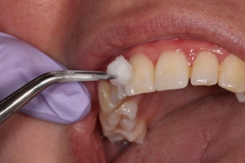
When evaluating the impact of a CO2 laser on vital human primary pulp, it becomes essential to closely examine key indicators that reflect pulp vitality. These indicators help determine the overall health of the tooth pulp and its response to dental treatments. Notably, **pain response**, **bleeding assessment**, and **temperature sensitivity** are crucial elements in assessing the state of the pulp. Observing these facets not only aids in immediate evaluations but also contributes to long-term dental care plans.
**Pain response** is a critical parameter used to gauge pulp vitality. During the evaluation, practitioners measure the pulp’s reaction to stimuli, whether thermal or mechanical. A vital, healthy pulp typically exhibits a transient pain response that dissipates quickly, while a compromised pulp may show prolonged or exaggerated pain. This is often evaluated using tools such as an electric pulp tester or a cold stimulus like ethyl chloride. Monitoring these reactions can help differentiate between a reversible pulpitis and more severe conditions.
Another significant aspect is the **bleeding assessment**. When a CO2 laser is used, it might create micro-incisions allowing for the observation of bleeding from the pulp. A healthy pulp shows a controlled bleeding response, which can be indicative of its vitality and overall health. Conversely, absent or excessive bleeding may signal an unhealthy pulp. Below is a simplified table demonstrating bleeding responses and their interpretations:
| Bleeding Response | Interpretation |
|---|---|
| Minimal | Potential Necrosis |
| Controlled | Healthy Pulp |
| Excessive | Possible Inflammation |
**Temperature sensitivity** is also a pivotal indicator in monitoring pulp vitality. Applying a cold or hot stimulus to the tooth can bring out vital reactions. In a vital pulp, the response to cold is sharp yet brief, while heat typically evokes a less intense reaction. In contrast, a non-vital pulp may not respond at all to temperature variations. This sensitivity testing, in tandem with other measurement techniques, provides a comprehensive view of pulp health, facilitating informed decisions about necessary treatment protocols.

CO2 Laser Treatment on Pulp: Evaluating Efficacy and Safety
The efficacy of **CO2 laser treatment** in the dental field has been a topic of substantial research, especially in terms of its impact on vital human primary pulp. Known for its precise cutting and coagulating properties, the CO2 laser has shown promise in reducing bacterial levels and ameliorating pulpal irritation. This section delves into various studies and findings that assert its reliability and safety.
- Minimized thermal damage
- Enhanced tissue healing
- Refined bacterial reduction capabilities
In comparative studies, traditional drilling methods have often come under scrutiny for causing micro-cracks and increased post-operative sensitivity. **CO2 lasers**, on the other hand, exhibit a minimally invasive approach, leading to better preservation of the pulp’s vitality. Detailed investigations show a remarkable reduction in bacterial counts post-treatment, making it a preferred choice for treating primary pulp.
| Treatment Method | Bacterial Reduction | Post-Operative Sensitivity |
|---|---|---|
| CO2 Laser | 90% | Low |
| Traditional Drill | 60% | High |
Another significant finding revolves around the **healing process** of the pulp post CO2 laser treatment. The laser’s ability to coagulate tissue instantly results in a reduced need for analgesics and faster wound healing. Moreover, the formation of a dentin bridge—an essential factor for the long-term viability of dental pulp—is notably more prominent when lasers are utilized compared to conventional methods.
Research continues to underscore the **safety aspects** of CO2 laser treatment. Unlike mechanical methods that can inadvertently enlarge the site of treatment and potentially expose the pulp to further infections, CO2 lasers offer a controlled and focused approach. This precision helps in maintaining the overall health of the dental structure while effectively targeting the problematic area, thereby establishing CO2 lasers as a safer alternative in contemporary dentistry.
Analyzing Short-Term and Long-Term Effects on Pulp Health
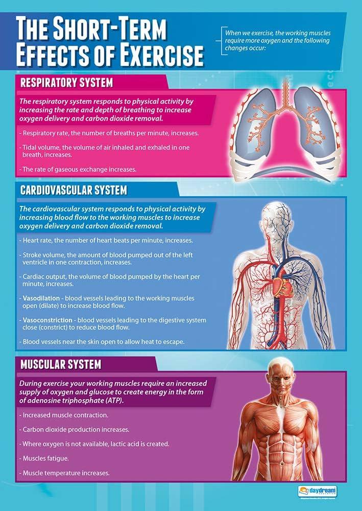
The application of CO2 lasers in dentistry has sparked interest due to their potential to offer precise and minimally invasive procedures. When it comes to assessing the impact on pulp health, it is essential to distinguish between the immediate and extended consequences. **Short-term effects** often include changes in pulp temperature and immediate cellular responses to laser exposure. The heat generated by the laser can cause a rise in pulp temperature, but modern CO2 lasers are designed to minimize this and reduce the risk of thermal damage.
- Immediate Inflammatory Response: Following laser exposure, the pulp may show an initial inflammatory reaction, which is a natural part of the healing process.
- Reduced Microbial Load: One of the notable short-term benefits is the reduction in microbial contamination, potentially decreasing the risk of postoperative infections.
**Long-term effects** are more complex, involving the pulp’s ability to repair and regenerate over time. Healing dynamics may include dentin bridge formation and the presence of fibrotic tissues. The pulp’s response to laser treatment is influenced by several factors, including laser parameters such as wavelength, power settings, and exposure duration. It’s also crucial to consider the variation in responses among different patients, which can be influenced by age, general health, and the current condition of the pulp.
| Short-Term Effects | Long-Term Effects |
|---|---|
| Temperature Control | Dentin Bridge Formation |
| Immediate Inflammation | Pulp Regeneration |
| Microbial Reduction | Longevity of Results |
A thorough understanding of these effects aids clinicians in making informed decisions about incorporating CO2 lasers into their practice. Continuous research and technological advancements are crucial in optimizing laser settings to enhance the benefits while minimizing potential drawbacks. while CO2 lasers offer promising outcomes for the health of vital human primary pulp, ongoing evaluation and customization of their use based on individual cases remain imperative.

Best Practices and Recommendations for Clinicians
To effectively evaluate the impact of CO2 laser therapy on vital human primary pulp, clinicians should incorporate several strategic practices into their protocols. **Accurate patient selection** is pivotal; prioritize patients with a clear medical history to avoid potential complications. Furthermore, utilize diagnostic imaging techniques like radiographs to assess the initial condition of the pulp tissue.
**Proper calibration of the CO2 laser** is another critical aspect. Ensure the power settings and exposure time are appropriate for the specific treatment being performed. Calibration minimizes the risk of thermal damage and enhances the precision of the procedure. Investing in **continuous training** for clinicians on the latest CO2 laser technology will contribute to more consistent and positive outcomes.
It’s equally important to apply **pre- and post-operative assessments**. Before the procedure, evaluate the vitality of the pulp using electric or thermal tests. After the treatment, monitor the healing progress, and be watchful for any signs of inflammation or infection. Regular follow-ups help in early identification of any complications and allow for timely interventions to preserve pulp vitality.
| Action | Purpose |
|---|---|
| Pre-operative Imaging | Assess initial pulp condition |
| Laser Calibration | Minimize thermal damage |
| Post-operative Monitoring | Ensure proper healing |
| Continuous Training | Update with latest technologies |
Utilizing **protective barriers** during the procedure can also enhance clinical efficacy. Use adjunctive materials like water spray or dental dams to further reduce heat dissipation and protect surrounding tissues. Clinicians should remain well-versed with both traditional and emerging studies, ensuring that the chosen practices are evidence-based and geared towards improving patient outcomes without compromising on safety.
Q&A
Q&A: Evaluating CO2 Laser Impact on Vital Human Primary Pulp
Q: What is the primary focus of the article?
A: The primary focus of the article is to evaluate the effects of CO2 laser treatment on the vital primary pulp tissue of human teeth. It explores the potential benefits and drawbacks of this technology in dental practices, particularly in pediatric dentistry.
Q: Why is CO2 laser treatment being considered for use on primary pulp tissue?
A: CO2 laser treatment is being considered due to its potential advantages, such as precise cutting, reduced bleeding, sterilization of the treated area, and quicker healing times. These factors could make it a valuable tool for managing the sensitive and delicate tissues in children’s teeth.
Q: How does CO2 laser treatment differ from traditional dental procedures?
A: CO2 laser treatment differs from traditional procedures primarily in its method of action. Instead of using mechanical drills, the laser uses a concentrated beam of light to remove or shape tissue. This method can be more precise and less invasive, reducing discomfort and improving recovery times.
Q: What were the key findings regarding the impact of CO2 laser on the vital pulp?
A: The key findings include observations on the laser’s ability to maintain pulp vitality, minimize inflammatory responses, and promote a favorable healing environment. However, the study also noted the importance of controlled laser settings to avoid thermal damage to the tissue.
Q: What methodologies were used in the study to evaluate the CO2 laser’s impact?
A: The study employed a combination of clinical observation, histological analysis, and various diagnostic tools to assess the pulp’s response to CO2 laser treatment. These methodologies helped to provide a comprehensive understanding of cellular and tissue-level changes post-treatment.
Q: Were any risks or potential disadvantages highlighted in the article?
A: Yes, the article highlighted several risks and potential disadvantages, such as the possibility of thermal damage if the laser parameters are not correctly set. It also mentioned the need for specialized training and equipment, which could be a barrier to widespread adoption.
Q: How does this article contribute to the field of pediatric dentistry?
A: This article contributes to pediatric dentistry by providing valuable insights into a relatively new technological application. It suggests that CO2 lasers could become a useful tool for managing primary pulp tissues, thereby potentially making dental treatments less traumatic for young patients.
Q: What future research directions does the article suggest?
A: The article suggests further research into optimizing laser settings, long-term effects of laser treatment on primary pulp, and comparative studies with other laser types and traditional methods. It also calls for larger sample sizes and diverse population studies to validate the findings.
Q: Who might benefit from reading this article?
A: Dentists, dental researchers, pediatric specialists, and students in dental programs would benefit from reading this article. It provides important information that could influence clinical practices and future research in the field of dental care for children.
Q: Is the CO2 laser ready for widespread use in pediatric dentistry based on this study’s findings?
A: While the study’s findings are promising, it concludes that CO2 laser should be used with caution and after thorough training. Further research and standardization of protocols are necessary before it can be recommended for widespread use.
By providing a thorough evaluation and shedding light on both the potential and limitations, this article serves as a valuable resource for anyone interested in the advancements of pediatric dental treatments.
In Conclusion
our exploration of the CO2 laser’s impact on vital human primary pulp shines a compelling light on the intersection of cutting-edge technology and classical dental science. As dental health continues to be a cornerstone of overall well-being, the potential of this laser technique to revolutionize treatment protocols cannot be overstated. Through meticulous research and careful consideration of both benefits and limitations, the path forward is illuminated with promise.
As with many pioneering endeavors, the journey is far from complete. The findings suggest a future ripe with possibilities but underscore the necessity for continued investigation and rigorous clinical trials. It is through this balanced lens of curiosity and caution that the true potential of CO2 lasers in preserving the vitality of primary pulp will be fully realized.
While we stand at the threshold of new horizons in dental therapeutics, one thing remains clear: the commitment to improving patient care through innovation and evidence-based practice is unwavering. As researchers, clinicians, and students of this ever-evolving field, we are called to press on, harvesting knowledge that will shape the smiles of tomorrow.


